Post a message
Replying to:
Merkel cell
Merkel cell were first identified by German anatomist Friedrich Sigmund Merkel in 1875 who, by using an osmium fixation and a silver staining, described them in the epidermal basal layer as large cells paler than keratinocytes and in close contact with enlarged terminal branches of myelinated afferent nerve fibers.
1. Distribution and localisation of Merkel cells
Merkel cells are epidermal cells localised in the basal layer of the epidermis and the epithelial sheath of hair follicles. The vast majority of Merkel cells are intimately associated with a nerve terminal but some are not. In all vertebrates, Merkel nerve endings are located in the basal layer of the epidermis, apart from birds, where they are located in the dermis. In mammals (apart from man), the largest accumulation of Merkel nerve endings is found in whiskers.
In adult human, the Merkel cell percentage vary from 0.5% to 5% in the epidermis but can vary in different areas of the body and during life. Merkel cells are more abundant in the glabrous regions of skin, lips, regions of the oral cavity, the erogenous zones where they are grouped in clusters (till 50 cells) around amyelinic nerve ends. They ar abundant in epidermal invaginations of the plantar foot surface called rete ridges. A sun-exposed skin can contain two times more Merkel cells than a non-exposed skin. These observations underline the occurrence of an undifferentiated cell pool able to regenerate Merkel cells during adult life; this would explain the regeneration of Merkel cells during cutaneous wound healing.
In hair follicles, they are present in the outer root sheath of the hair follicle and the bulge which is a stem cell reservoir. They are also numerous in specialized epithelial structures of the hairy skin called touch domes. The Merkel cell density in hair follicles change along the hair cycle with a maximum during the anagen phase and a minimum during the catagen and telogen phases
2. Morphology of Merkel cells
At the ultrastructural level, they show a lobulated nucleus, finger-like protoplasmic protusions in the part of the cell opposite to the nerve terminal, relatively clear cytoplasm containing round 80-160 nm neurosecretory granules with a central electron-dense core surrounded by a clear halo and a single limiting membrane and which are facing sensory neurones. They project cytoplasmic spines to neighbouring keratinocytes with which they are connected by desmosomes .
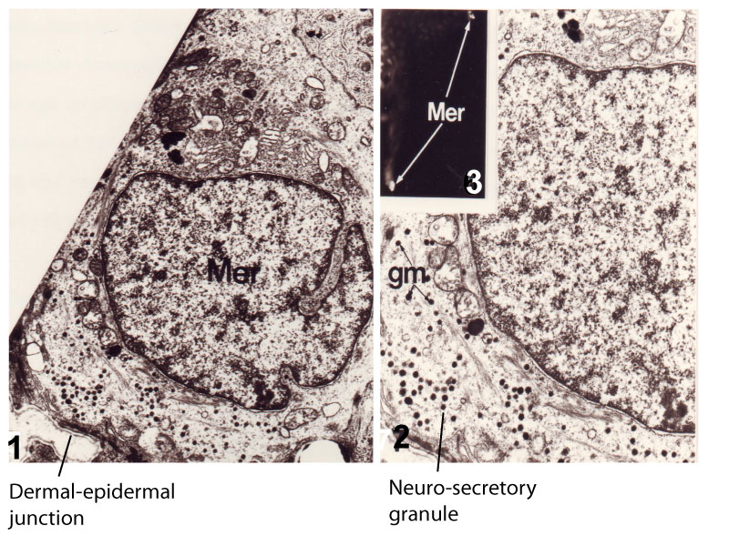
- Merkel cell observed by transmission electron microscopy in the epidermis of human skin transplanted onto the nude mouse, two months after grafting (X 10000); 2) detail of figure 1 (X 20000); 3) two Merkel cells labelled by indirect immunofluorescence with Troma 1 antibody in a human epidermis.
- Note the presence of neuro-secretory granules at the basal pole of the cell, of the Golgi apparatus at the apical pole, and the occurence of desmosomes with neighbouring keratinocytes.
Merkel cells can be detected on histological sections with uranaffin stains and in immunohistology with antibodies against various specific antigens including keratin n° 18, 19, or 20, neuron-specific enolase (glycolytic enzyme of neuronal/neuroendocrine cells), chromogranin (68 kDa protein component of neurosecretory granules), synaptophysin (acid 38 kDa protein associated to synaptic vesicles), neural cell adhesion molecule (N-CAM) and various neuropeptides (protein gene product 9.5, met-enkephalin, CGRP , VIP ).
3. Origin of Merkel cells
Whether Merkel cells originate from embryonic epidermal or neural crest progenitors has been a matter of intense controversy since their discovery in 1875. Moreover, how Merkel cells are maintained during adulthood was till recently unknown.
One hypothesis posited that Merkel cells derive from neural crest cells since they are excitable cells that synthesize neuropeptides and express presynaptic molecules and proneural transcription factors like many other neural crest-derived cells. Moreover, results from lineage experiments in quails and in mice suggested that Merkel cells originate from neural crest stem cells.
Another hypothesis was that Merkel cells originate from epidermal progenitors since they are localised in the basal layer of the epidermis and express keratins (K8, K18, K19, and K20). Moreover they are present in the epidermis before the appearance of nerves. In humans, they are identifiable and transplantable, several weeks before nerves reach the fetal epidermis.
Studies reporting lineage-tracing experiments with transgenic mice have been recently published and appears to clarify the question. Atoh1/Math 1 is expressed in developing and adult Merkel cells . (K14; Atoh1-CKO) or (Wnt1; Atoh1-CKO) mice have been used to study the results of conditional deletion of the Atoh1 from the skin or neural crest lineages, respectively (Morrisson et al., 2009; Van Keymeulen et al., 2009). Deletion of Atoh1 from the skin lineage results in the absence of Merkel cells in all skin locations including the whisker region while deletion from the neural crest lineage had no effect on this cell population. These recent data demonstrate an epidermal origin of mammalian Merkel cells. In adults, Merkel cells undergo slow turnover and are replaced by cells originating from epidermal stem cells, not through the proliferation of differentiated Merkel cells.
4. Merkel cell functions
For more than a century, neurobiologists have postulated that Merkel cells act as slow-adapting type I mechanoreceptor and are responsible for the specialized coding properties that allow their afferent nerves to resolve fine spatial details. Merkel cells send dendritic processes between keratinocytes and Langerhans cells and can form close association with type A-beta sensory neurones to form the Merkel cell-neurite complex which is among the most sensitive touch receptors mediating one form of light touch important for tactile two-point discrimination and for detection of shapes, curvature and textures.
A recent genetic knockout approach has allowed Maricich et al. (2009)to demonstrate that Merkel cells are essential for these responses and could represent the sensory receptor cells of the complexes. For this purpose, conditional knockout Atoh1-CKO mice have been engineered. As already said, Atoh1 is a transcription factor expressed by Merkel cells in all areas of the skin. In this study, Hoxb1Cre allele, which is expressed throughout the dermis and epidermis of body skin, but not head skin, has been used to delete Atoh1 from the body skin and foot pads of this transgenic mice. As expected, Merkel cells are absent from these areas in Atoh1CKO animals. Ex vivoskin/nerve preparations from Atoh1CKO animals demonstrate complete loss of the characteristic neurophysiologic responses normally mediated by Merkel cell-neurite complexes. Merkel cells are, therefore, required for the proper encoding of Merkel receptor responses, suggesting that these cells form an indispensible part of the somatosensory system.
If Merkel cells are sensory receptor cells, then they must transmit signals through synaptic contacts with somatosensory neurons. Consistent with this notion, Merkel cells contain dense-core vesicles that resemble neurosecretory vesicles. Moreover, Merkel cell-neurite complexes have membrane densities like those at synaptic active zones. Haeberle et al.(2004) have purified Merkel cells from touch domes and used DNA microarrays to compare gene expression in Merkel cells and other epidermal cells. They identified 362 Merkel-cell-enriched transcripts, including neuronal transcription factors, presynaptic molecules, and ion-channel subunits. By immunohistology, they showed that Merkel cells express presynaptic active-zone constituents including the active-zone-matrix protein Piccolo, Rab3C, vesicular glutamate transporter 2, and cholecystokinin 26-33, synaptic vesicle proteins, and molecules required for neuropeptide production and glutamate release. Moreover, live-cell imaging experiments revealed that Merkel cells have functional L- and P/Q-type voltage-gated Ca2+ channels which are channels essential for synaptic transmission. Together, their data demonstrate that Merkel cells are excitable cells and designate glutamate and CCK8 as candidate neurotransmitters at synapses between Merkel cells and sensory afferents in vivo. This conclusion was strengthened by the abundance of neuronal transcription factors (including Math1 and Gfi1) found to be enriched in Merkel cells.
The Merkel cell population is heterogeneous. Merkel cells are not all associated with nerves and Merkel cells of different locations of the body express different neural (neurofilament proteins, nerve growth factor receptor, synaptophysin) and epithelial (villin) proteins. These findings led to the suggestion that while some Merkel cells function as mechanoreceptors of tactile stimuli, other “Merkel-like” cells, with similar appearance as Merkel cells, but without contact to nerve terminals, form part of the diffuse neuroendocrine system involved with modulation of peripheral neural responses. It is these cells, rather than those acting as mechanoreceptors, that are believed to be at the origin of a highly malignant skin cancer called Merkel cell carcinoma.
Bibliography
Merkel, F., Tastzellen and Tastkoerperchen bei den Hausthieren und beim Menschen. Arch Mikrosc Anat, 1875. 11: p. 636-652.
Maricich S. et al. Merkel Cells Are Essential for Light-Touch Responses(2009). Science 324 : 1580-1582.
K. M. Morrison, G. R. Miesegaes, E. A. Lumpkin, and S. M. Maricich.
Mammalian Merkel cells are descended from the epidermal
lineage (2009). Dev Biol., 336(1): 76–83.
A. Van Keymeulen, G. Mascre, K. K. Youseff, I. Harel, C. Michaux, N. De Geest, C. Szpalski, Y. Achouri, W. Bloch, B. A. Hassan, et al. (2009) ;Epidermal progenitors give rise to Merkel cells during embryonic development and adult homeostasis. J. Cell Biol. :187, 91-100.
Site powered by SPIP 3.0.17 + AHUNTSIC

Visitors logged in: 48